The Power of Regenerative Medicine for Neurological Conditions #
NeuroCytonix is pioneering the future of brain repair through regenerative medicine, a branch of biomedical science focused on harnessing the body's regenerative capabilities to repair or replace damaged tissues and organs.
Regenerative medicine explores methods to activate the growth and repair of neural cells and tissues affected by injury or disease. This field encompasses a range of approaches, including stem cell therapy, tissue engineering, and the use of growth factors and biomaterials though none have achieved success to date.
Researchers have been developing protocols to encourage neuroregeneration, aiming to restore lost functions and potentially reverse neurological disorders. Regenerative medicine holds promise for revolutionizing the treatment of brain-related conditions and improving the quality of life for those affected.
Our Groundbreaking Neural Regeneration Technology #
Our cutting-edge technology is designed to harness the body’s natural cell signaling pathways to restore the nervous system in people affected by neurological conditions.
Our technology uses non-invasive functional neurogenesis stimulation to help facilitate repairing the circuitry of the brain. Functional neurogenesis stimulation plays an important role in neural tissue engineering, leveraging the body's natural signaling mechanisms to promote the development and functionality of neural tissues. The goal is to stimulate the brain’s ability to heal itself, by guiding the growth, organization, and repair of neural networks.
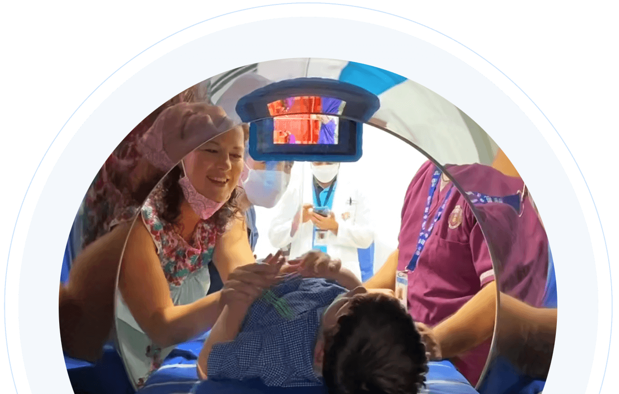
How does our technology work? #
Our device of functional neurogenesis stimulation has rings of electromagnetic signal emitters that direct radiofrequency waves to affected areas of the brain. Our doctors can target either one specific area or multiple areas in the body, depending on where the nervous system is damaged, and the type of condition being addressed.
The device emits radiofrequency waves, which are a low-energy form of electromagnetic radiation. Sensors that generate magnetic fields and radiofrequency waves are positioned on the inner walls of the device. A Magnetic Resonance Imaging (MRI) scan is conducted to visualize the brain structure and precisely locate the area where the radiofrequency signals should be focused.
Our neurologists study the MRI scan data to customize the protocol based on each individual’s neurological condition.
Our Regenerative Medicine Research #
With the nature of our approach, research on our protocol’s impact on neurological disorders is ongoing. Evidence from our imaging data suggests there is an increase in gray and white matter volume, both essential components of brain structure, along with a rise in the number of neural tracts—pathways that connect different regions of the brain.
Our research suggests that the electromagnetic signals may activate the brain's natural repair mechanisms, encouraging the regeneration of damaged cells.
Published research demonstrates the potential of tissue engineering technology to repair cartilage and promote brain recovery. Scientific studies show that this technology can trigger chondrogenesis—the process of cartilage formation—by stimulating specific proteins that promote the growth of chondrocytes, the cells responsible for healthy cartilage. This breakthrough offers hope for developing treatments for degenerative conditions such as osteoarthritis, which damage joints over time.
Furthermore, our approach may also support neurogenesis—the growth and repair of nerve cells in the brain—by activating well-documented proteins and neuronal growth factors. These factors play a critical role in strengthening neural connections and restoring the brain’s network, potentially offering new ways to repair damage caused by incurable neurological disorders, such as cerebral palsy, autism and traumatic brain injury.
How are our protocols administered? #
Prior to administering the protocol, each medical case is reviewed by our team of neurologists to determine eligibility. An MRI scan is used to visualize the brain structure and precisely locate the area to focus the radiofrequency signals. The MRI data is then analyzed to customize the protocol based on the individual's neurological condition. Our technicians then calibrate the device to administer calculated doses of radiofrequency waves during a one-hour session, conducted at the same time each day, for 28 days.
How We Study Our Technology’s Impact #
Over 500 people with various neurological conditions have now received our protocols at our clinical research center in Monterrey, Mexico. Encouraged by results from our initial clinical trial with children suffering from cerebral palsy, we have expanded our trials to assess the impact of our protocols on autism spectrum disorders and have also developed research protocols for stroke, traumatic brain injury (TBI), vascular dementia, and the neurological impacts of COVID-19.
NeuroCytonix Mexico Clinical Research Center #
Discover the future of medical research at our state-of-the-art clinical research center.